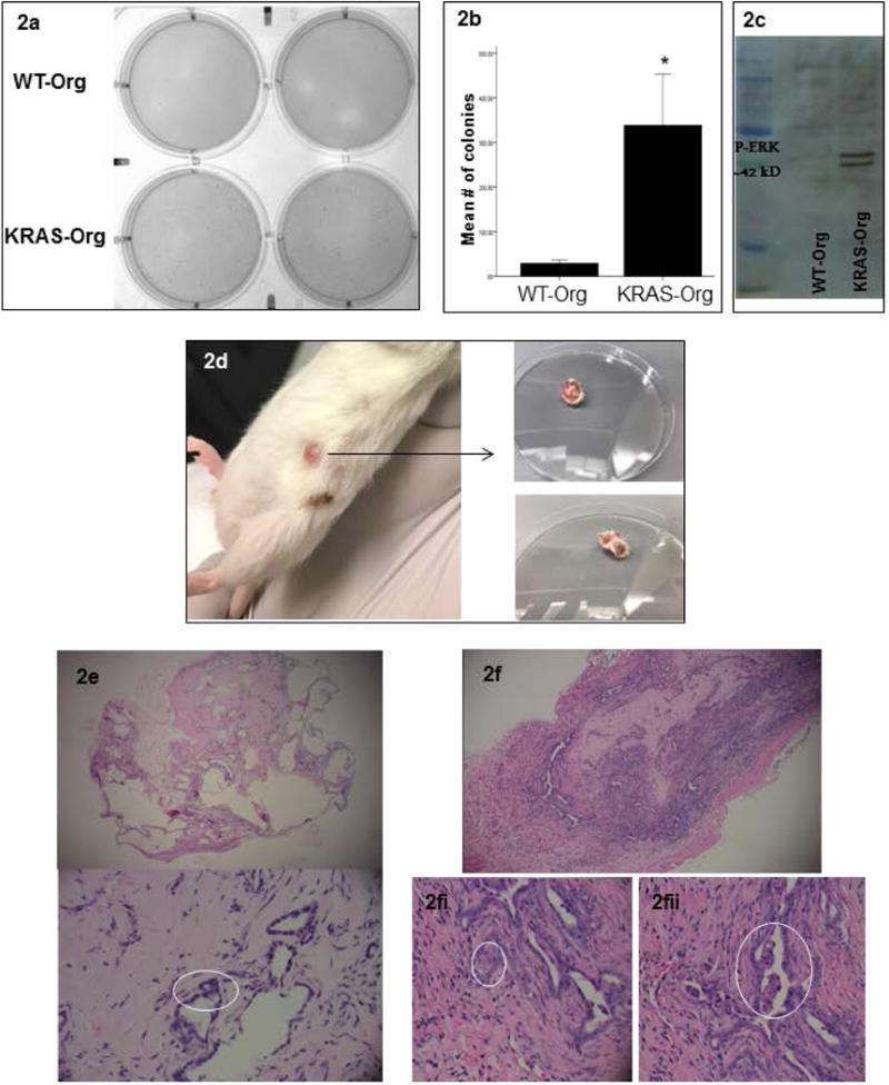
Figure 2.

KRAS mutations in pancreas organoids induce cancer-related phenotypes in vitro and in vivo; (a) representative of a colony formation assay comparing WT and mutated KRAS carrying organoids; (b) quantification of colonies formed by WT and mutated KRAS carrying organoids, bar graphs show means ± SE of n=3 experiments, asterisk indicates p<0.05; (c) representative of a p-ERK western blot in the WT vs. mutated KRAS carrying organoids’ lysates; (d) KRAS carrying organoids formed tumors in NSG mice 10 weeks after subcutaneous injection; right, tumors after resection; (e) histology of the tissue formed 12 weeks after WT organoids injection: Top panel is a 4× view showing benign pancreatic ductal structures with variable degrees of dilatation. No internal papillary formations are seen. Bottom panel is a 10× view of benign ductal structures. Nuclei are small and bland (circled). (f) 4× view of a tumor formed from KRAS carrying organoids in NGS mice after 10 weeks showing back-to-back gland formation, densely cellular ductal structures with infiltrative edges representing an early invasive phenotype; (2.f.i) 10× view of an area of cells with large nuclei that are not uniformly bland, showing noticeable nucleoli; (2.f.ii) 10× view of luminal papillary structures, reminiscent of pancreatic intraepithelial neoplasia lesions.
