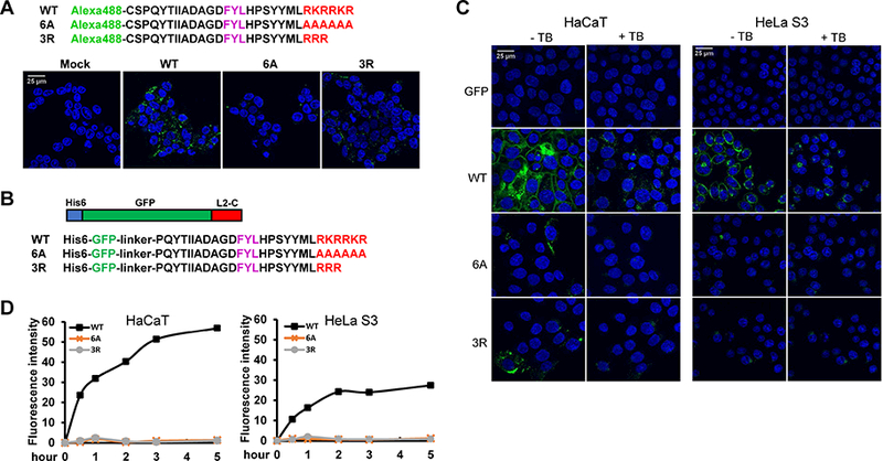
Figure 2. The L2 basic sequence displays cell-penetrating activity.

(A) Amino acid sequence of HPV16 L2 C-terminal peptides containing the wild-type (WT) basic sequence or the six alanine (6A) or three arginine (3R) mutations conjugated to Alexa Fluor 488. 293T cells were mock-treated or incubated with fluorescent peptides for three h at 37°C and examined by confocal microscopy. (B) Schematic diagram and C-terminal sequences of GFP-L2 fusion proteins. (C) HaCaT and HeLa S3 cells were incubated with GFP-L2 fusion proteins for three h. Cells were examined by confocal microscopy, then treated with trypan blue (+TB) to quench extracellular fluorescence, and the same fields were re-imaged. (D) HeLa S3 and HaCaT cells were incubated with GFP-L2 fusion proteins for various times. Cells were then treated with trypsin, and fluorescence was measured by flow cytometry. Mean fluorescent intensity (MFI) was plotted at the indicated time periods.
