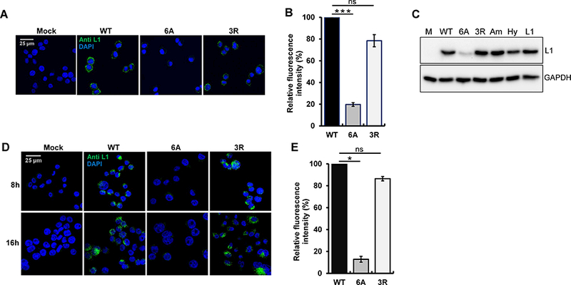
Figure 3. The basic region of HPV16 L2 is not essential for virus binding and internalization.

(A) HeLa-sen2 cells were mock-infected or incubated at 4°C for two h at MOI of 20 with wild-type, 6A, or 3R HPV16 PsV. Non-permeabilized cells were stained with anti-L1 antibody (green) and examined by confocal microscopy. (B) HeLa-sen2 cells were treated as described in panel A, detached with EDTA, and stained with anti-L1 antibody. MFI of the cells was measured by flow cytometry and normalized to cells incubated with wild-type HPV16 PsV. Mean results and standard deviation are shown (n=3). ***p<0.001; ns, not significant. (C) HeLa-sen2 cells were treated as described in panel A with the following PsVs: wild-type HPV16 (WT), 6A mutant, 3R mutant, mutant with amphipathic CPP (Am), mutant with hydrophobic CPP (Hy), and L1-only PsV (L1). After two h at 4ºC, cells were washed, lysed, and bound virus was assessed by SDS-PAGE and blotting for L1 (top panel). GAPDH is a loading control (bottom panel). (D) HeLa cells-sen2 were mock-infected or infected at MOI of 50 at 4°C for two h with wild-type, 6A or 3R HPV16 PsV and then washed and shifted to 37°C for eight or 16 h to allow internalization. Permeabilized cells were stained with anti-L1 antibody (green) and examined by confocal microscopy. (E) HeLa-sen2 cells were infected as described in panel D and harvested by trypsinization 6 h after shift to 37ºC. Permeabilized cells were stained with anti-L1 antibody and analyzed by flow cytometry. MFI of the cell populations was normalized to cells infected with wild-type HPV16 PsV. Mean results and standard deviation are shown (n=3). *p<0.05. See also Figure S4.
