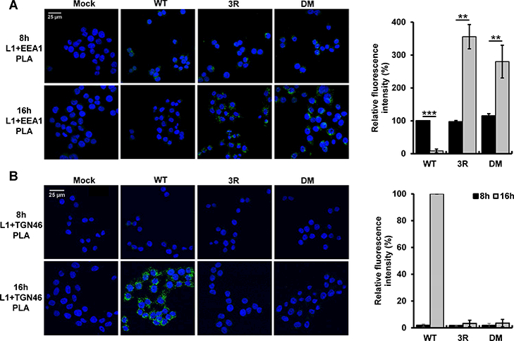
Figure 4. The 3R mutant accumulates in endosomes.

HeLa-sen2 cells were mock-infected or infected at 37ºC at MOI of 100 with wild-type, 3R or retromer binding site (DM) mutant HPV16 PsV. At eight and 16 hpi, PLA was performed with anti-L1 antibody and either an EEA1 (panel A) or a TGN46 (panel B) antibody. PLA signal is green. Multiple images obtained as in left panels were processed with BlobFinder software to determine the fluorescence intensity per cell. The graphs in the right panels show mean results and standard deviation (n=3), in which the results for the EEA1/L1 samples and the TGN46/L1 samples were normalized to those of cells infected with wild-type HPV16 PsV at eight and 16 hpi, respectively. Black bars, 8 hpi; grey bars, 16 hpi **p<0.01; ***p<0.001.
