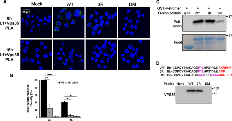
Figure 5. The 3R mutant is defective in accessing retromer during HPV infection.

(A) HeLa-sen2 cells were infected as in Figure 4. At eight and 16 hpi, PLA was performed with anti-L1 antibody and an antibody recognizing Vps35 to assess L1 in proximity to retromer. (B) Multiple images obtained as in panel A were processed and displayed as in Figure 4 with the mutant samples normalized to that of cells infected with wild-type PsV at eight hpi. Black bars, wild-type; dark grey bars, 3R mutant; light grey bars, DM mutant. *p<0.05; **p<0.01; ***p<0.001. (C) GST-tagged retromer adsorbed to glutathione beads was incubated with GFP-L2 fusion proteins. After pull-down, proteins bound to retromer were separated by SDS-PAGE and detected by blotting with anti-GFP antibody (top panel). Bottom panel shows input GFP-L2 fusion proteins. (D) Top. Sequences of wild-type and mutant biotinylated peptides. Bottom. Biotinylated peptides were incubated with extracts of uninfected HeLa S3 cells, pulled-down with streptavidin, and separated by SDS-PAGE. Retromer associated with the peptide was detected by blotting for Vps35.
