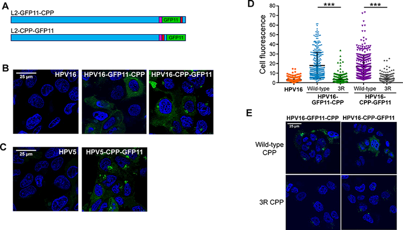
Figure 7. Reconstitution of split GFP by infection with PsV containing L2-GFP11.

(A) Schematic diagrams of L2 protein with tandem GFP11 (green) inserted upstream of RKRRKR (L2-GFP11-CPP) or at the C-terminus of L2 (L2-CPP-GFP11). The bulk of the L2 protein is shown in blue; CPP in red; and retromer binding sites in purple. See Figure S3A for sequences. (B) Clonal HaCaT/GFP1–10NES cells were infected at MOI of 2000 with untagged HPV16 PsV or with a PsV containing GFP11-tagged L2. Three hpi, cells were examined by confocal microscopy for GFP fluorescence. (C) As in panel (B), except cells were infected with HPV5 PsV containing untagged or GFP11-tagged L2. (D) Fluorescence of cells in images as in panel B was quantified for ~250 cells for each condition. Results show the fluorescence intensity of individual cells plotted in arbitrary units from three independent experiments. ***p<0.0001. (E) Cells were infected as described in panel B with GFP11-tagged PsV containing the wild-type or the 3R mutant CPP. Three hpi, cells were examined by confocal microscopy for GFP fluorescence. See Figures S2 to S5.
