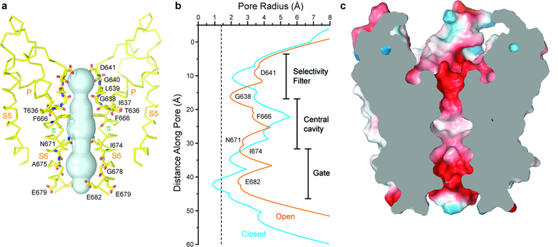
Figure 4: Open pore of TRPV3.

a, Pore-forming domain of TRPV3(Y564A)2-APB with residues lining the pore shown as sticks and the pore profile shown as a space-filling model (cyan). b, Pore radius calculated using HOLE54 for TRPV3 (blue) and TRPV3(Y564A)2-APB (orange). The vertical dashed line denotes the radius of a water molecule, 1.4 Å. c, Coronal section of the TRPV3(Y564A)2-APB pore-forming domain, with surface coloured by electrostatic potential.
