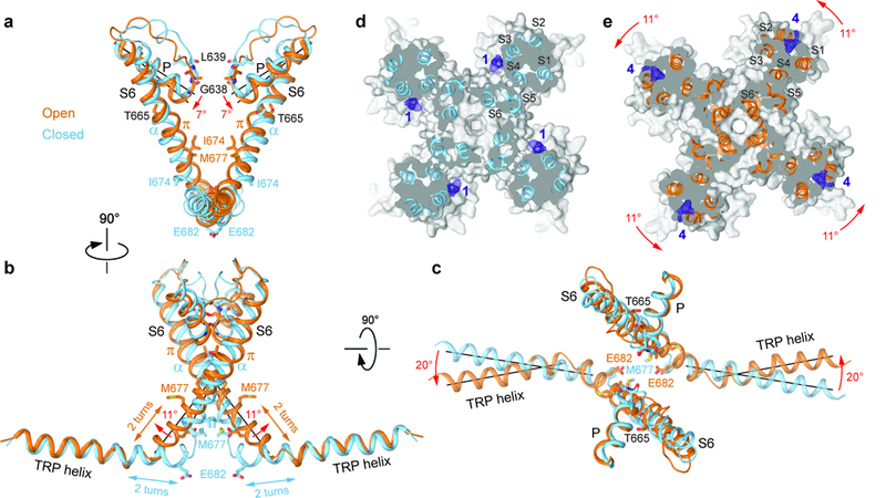
Figure 5: Structural changes associated with TRPV3 channel opening.

a-c, Superposition of the P-loop, S6 and TRP helices of TRPV3 (blue) and TRPV3(Y564A)2-APB (orange) viewed parallel to the membrane (a,b) or extracellulary (c). Only two of four subunits are shown for clarity. Residues lining the pore are shown as sticks. Rotations of the pore, S6 and TRP helices in TRPV3(Y564A)2-APB relative to TRPV3 are indicated by red arrows. d-e, Sections through the space filling models of TRPV3 (d, blue) and TRPV3(Y564A)2-APB (e, orange) made perpendicular to the pore axis and viewed extracellularly. Non-protein densities at site 1 (d) and site 4 (e) are shown as purple mesh at 4σ. The structures are aligned based on their pore domains. The 11° rotation of the S1-S4 domains in TRPV3(Y564A)2-APB relative to their position in TRPV3 is indicated by the red arrows.
