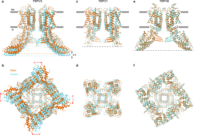
Figure 6: Comparison of gating rearrangements in different TRPV channels.

a-f, Superposition of structures in the open (orange) and closed (blue) states for TRPV3 (a-b, TRPV3(Y564A)2-APB and TRPV3, respectively), TRPV1 (c-d, PDB: 5IRX and PDB: 5IRZ, respectively) and TRPV6 (e-f, PDB: 6BO8 and PDB: 6BOA, respectively) viewed parallel to the membrane (a,c,e) or intracellularly (b,d,f). Only two of four subunits are shown in (a,c,e), with the front and back subunits omitted for clarity. The structures are aligned based on their pore domains. Note, the TRPV3 structure becomes shorter (black arrows) and its intracellular skirt undergoes substantial rotation (red arrows) during channel opening, while the overall architecture of TRPV1 and TRPV6 remains the same.
