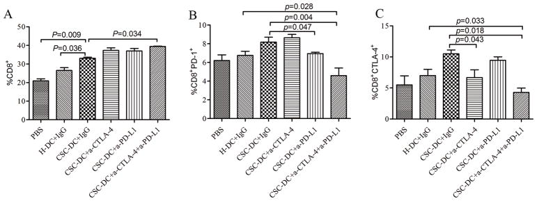
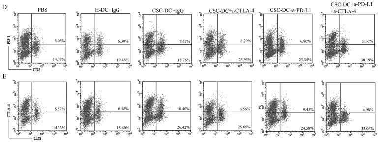
Fig. 3.
CSC-DC vaccination combined with immune checkpoints blockade changed the phenotype of CD8+ T cells. A, B, C: The percentage of CD8+ T cells (A), PD-1+CD8+ T cells (B) and CTLA-4+CD8+ T cells (C) in the spleens harvested from mice treated with PBS, H-DC vaccines, CSC-DC vaccines alone or in combination with anti-CTLA-4 and/or anti-PD-L1 as indicated. Each column represents the mean±SE of three independent experiments performed. D, E: FACS plots representing PD-1+CD8+ T cell (D) and CTLA-4+CD8+ T (E) populations in the spleens of mice treated as indicated. Single-cell suspensions were prepared from spleens on day 30 after tumor challenge and stained for immune checkpoints, PD-1 and CTLA-4.


