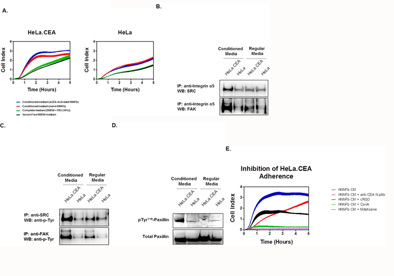
Figure 4. Medium from sCEA-activated fibroblasts preferentially augments the adherence of CEA-expressing cells.

A. sCEA-activated fibroblasts promote the adherence of CEA-expressing HeLa (HeLa.CEA) cells, but not of parental HeLa cells. Cells (2.0 × 10⁴) were dispensed to E-plate wells pre-treated with serum-free, complete medium (negative control) or medium recovered from either naïve or sCEA-activated fibroblasts. Cellular adhesion is reported as averaged Cell Index (CI) values (± SEM). B. Incubation of CEA-expressing cells in fibroblast-conditioned media promotes integrin signalling and recruitment of FAK and SRC. Suspensions of HeLa or HeLa.CEA (5 × 10⁶) cells were incubated in either regular medium or fibroblast-conditioned medium for 30 minutes. Adhering cells were lysed in RIPA buffer followed by immuno-precipitation using an anti-human integrin α5 mAb and immuno-blotting for either SRC or FAK. C. Incubation of CEA-expressing cells in fibroblast-conditioned medium promotes FAK and SRC phosphorylation. Lysates of adhering cells were immuno-precipitated using anti-human FAK or SRC mAb and immuno-blotted for phospho-tyrosine using mAb 4G10. D. Incubation of CEA-expressing cells in fibroblast-conditioned medium favors a substantial increase in paxillin phosphorylation. Lysates of adhering cells were immuno- blotted for both total and phospho-paxillin. E. Disruption of the Fn-CEA/integrin interactions inhibits cellular adherence. Addition of cRGD, anti-CEA N pAb, miltefosine or ConA to adhering HeLa.CEA cells reduces cellular adhesion. HeLa cells (2.0 × 10⁴) were dispensed to E-plate wells containing medium from either naïve or sCEA-activated fibroblasts. The impact of adding these inhibitors was monitored in real time as changes in relative electrical impedance (CI) at 1-min intervals, for 5 hours.
