Figure 3.
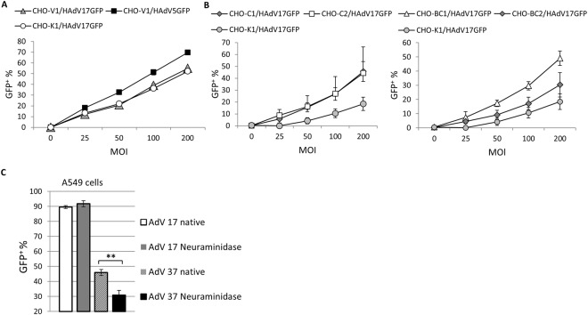
HAdV17 receptor usage. (A) GFP positive CHO-V1 cells (CAR positive) determined by flow cytometry after transduction with HAdV5GFP and HAdV17GFP at increasing MOIs as indicated. Percentage provided indicates % of GFP-positive cells. (B) Percentage of GFP-positive cells in transduced CD46 positive CHO-C2, CHO-C1, CHO-BC1, CHO-BC2 cells determined by flow cytometry after transduction with HAdV17GFP at increasing MOIs. CHO-K1 cells were used as controls. (C) Neuraminidase treated and untreated (native) A549 cells were infected with HAdV17 and HAdV37 and the percentage of GFP positive cells was determined. **P < 0.01.

