Figure 7.
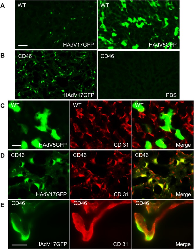
Immunofluorescence analysis of liver sections. Mice were sacrificed 3 days after vector injection. (A) HAdV17GFP injection induced weak GFP signals in liver sections of C57BL/6 wild type (WT) mice (left), comparing to HAdV5GFP injection (right). (B) HAdV17GFP injection induced high GFP signals in liver sections of CD46 transgenic mice (left), but no detectable signal after PBS injection (right). (C) There are no obvious colocalization of GFP and CD31 in liver sections of WT mice after HAdV5GFP injection. (D) GFP signals were highly colocalized with CD31 in liver sections of CD46 transgenic mice injected with HAdV17GFP. (E) High magnification of GFP signals with CD31 in liver sections of WT mice after HAdV5GFP injection. Scale bars: A-B, 50 µm; C-D, 20 µm; E, 25 µm.

