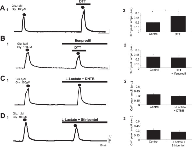
Figure 3.
L-Lactate-induced potentiation depends on the redox state of the cell. (A1,B1,C1 and D1) Intracellular Ca2+ signals (average Fura-2 ratio) following 2 successive co-applications of glutamate/glycine (2 min; dots) and recorded from 4 different cultures in presence of DTT (1 mM) (ncell = 43; A1), DTT (1 mM) + Ifenprodil (2 μM) (ncell = 21; B1), L-Lactate (10 mM) + DTNB (200 μM) (ncell = 62; C1) or L-Lactate (10 mM) + Stiripentol (200 μM) (ncell = 42; D1). Only the reducing agent DTT alone potentiates the Ca2+ response evoked by the co-application of glutamate/glycine (A1). (A2,B2,C2 and D2) Bar charts summarizing the significant potentiating effect of DTT (ncult = 9; ncells = 343; A2) blocked by Ifenprodil (ncult = 8; ncells = 301; B2) and the blockade of the L-Lactate-induced potentiation in presence of DTNB (ncult = 8; ncells = 407; C2) or Stiripentol (ncult = 9; ncells = 298; D2) on the Ca2+ signal triggered by co-application of glutamate/glycine. Results are presented as means ± SEM (*p < 0.05; paired t-test).

