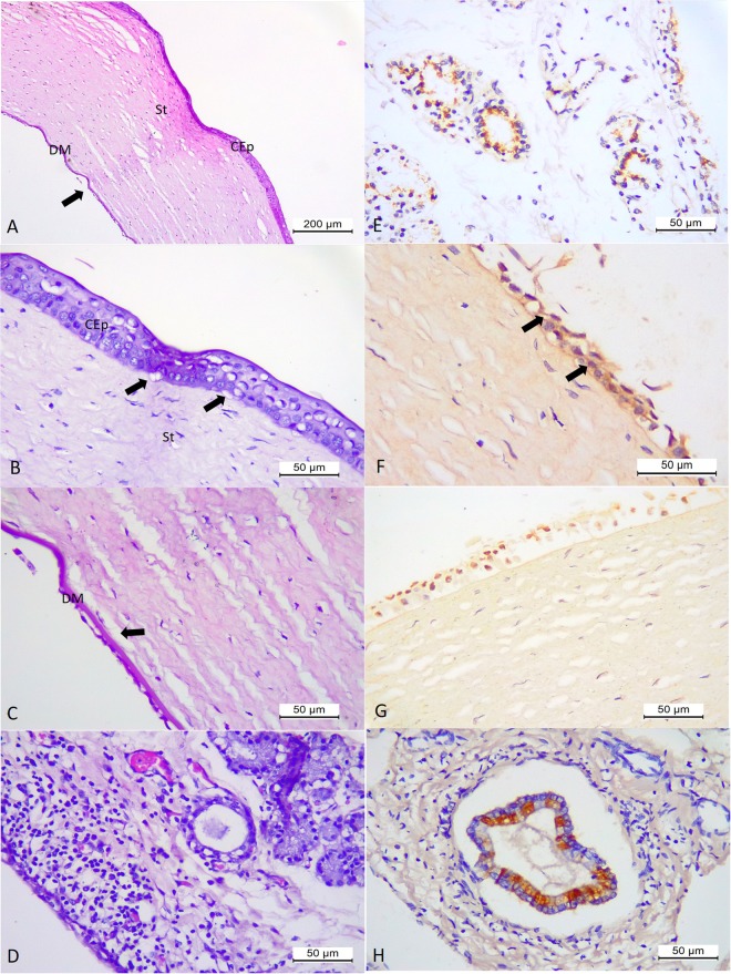
Figure 6.
Histopathologic and immunohistochemical findings associated with CAdV-1 infection in puppy # 15. Eye, there is marked oedema of the stroma (St) and dislocation (arrow) of the Descemet’s membrane (DM) of the cornea (A). Eye, observe ballooning degeneration of the epithelial cells of the corneal anterior (CEp) with disruption (arrow) of the basement membrane (B) and at Descemet’s membrane (DM) of the cornea, and without any inflammatory reaction at the corneal endothelium (B,C). Conjunctiva, observe focus of moderate lymphoplasmacytic inflammatory cells that are adjacent to degenerative and necrotic epithelial cells of the lacrimal gland (D). There is the positive immunoreactivity to CAdV-1 antigens at the degenerated and necrotic epithelial cells of the lacrimal glands at the conjunctiva (E), the degenerated epithelial cells (arrows) of the cornea (F,G), and bile duct epithelial cells of the liver (F). (A–D) Haematoxylin and Eosin stain; (E–H) immunoperoxidase. Bar, (A) 200 µm, (B–G) 50 µm.

