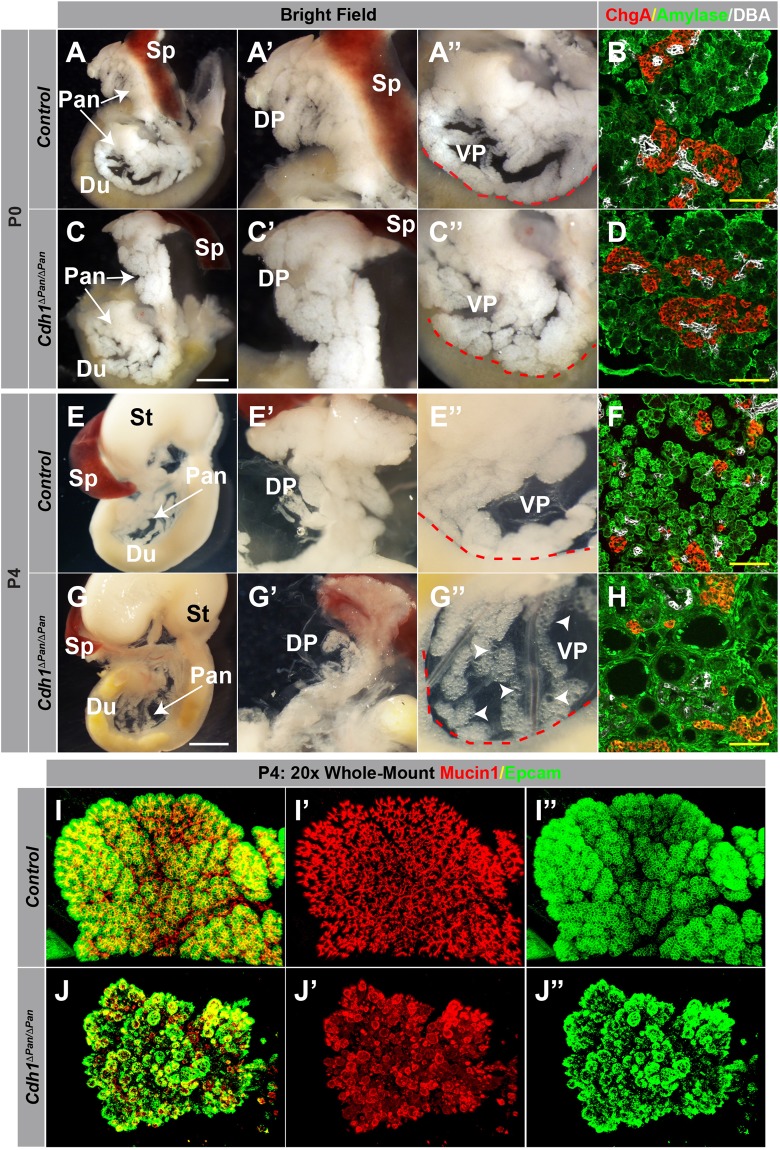
Figure 2.
E-cadherin plays an indispensable role in postnatal exocrine tissue architecture. (A,C,E,G) Whole-mount images of the pancreas (Pan), spleen (Sp), stomach (St) and duodenum (Du) of control and Cdh1ΔPan/ΔPan animals at P0 (A,C) and P4 (E,G). Higher magnification views of the dorsal (DP, A’,C’,E’,G’) and ventral (VP, A”,C”,E”,G”) pancreas are shown. White scale bars = 250 μm. Red contour indicates edge of duodenum. White arrowheads indicate cystic exocrine structures. (B,D,F,H) Immunofluorescence analysis of acinar (amylase, green), ductal (DBA, white), and endocrine (Chromogranin A, red) compartments of control (B,F) and Cdh1ΔPan/ΔPan (C,H) mice at both P0 and P4. Yellow scale bars = 100 μm. (I,J) Whole-mount immunofluorescence images showing the distribution of ductal (Muc1, red) and exocrine (Epcam, green) compartments in the control (I) or Cdh1ΔPan/ΔPan (J) pancreas at P4.

