Figure 4.
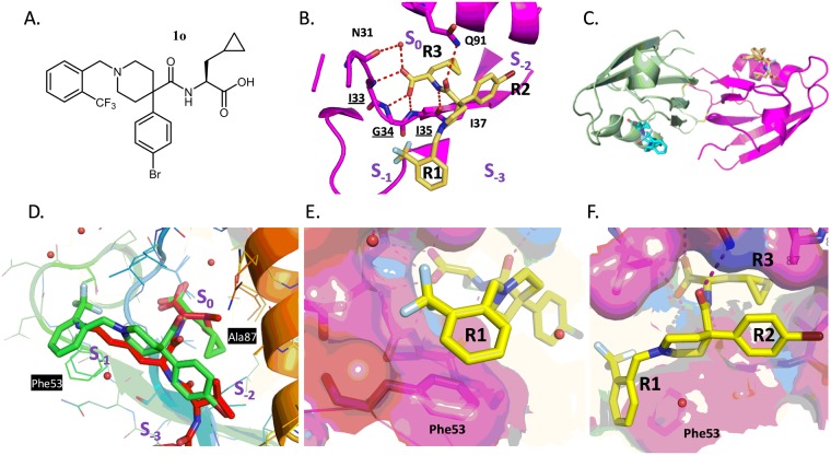
Structure of PICK1 with compound 1o. (A) Structure of compound 1o. (B) Specific binding of 1o to the PICK1 PDZ domain showing a 3-pronged pharmacophore entering pockets S0 (“R3”), S−1 (“R1”), and S−2 (“R2”). (C) Overall conformation of the PICK1 dimer and location of compound 1o binding (yellow and blue) in the peptide groove of each monomer (green and magenta). (D) Superposition of PICK1 alpha-carbons with the 3HPK PICK1 structure highlighting difference between peptide (SVKI; Red) and compound 1o (green) (E), Stacking between R1 aromatic group of compound 1o and Phe53 including the subpocket (F), Surface representation demonstrating interaction between fluorophenyl (R2) and Ala87, Lys83.

