Figure 7.
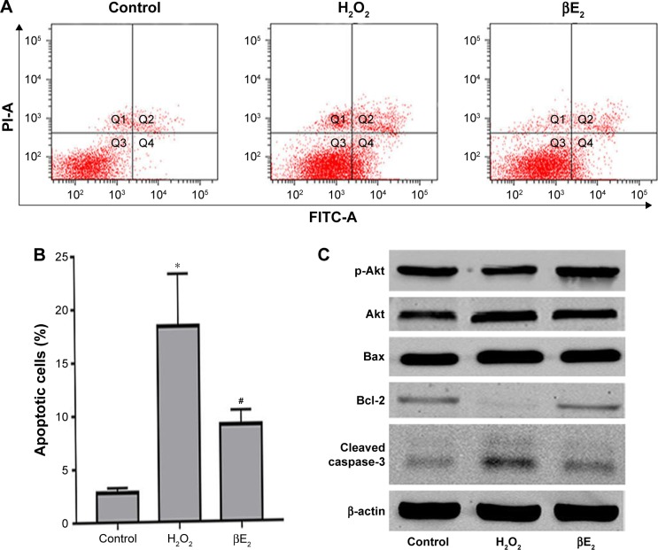
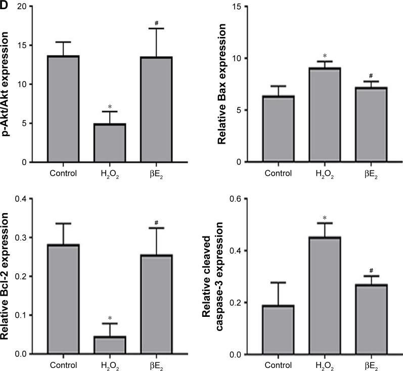
βE2 treatment decreases H2O2-induced apoptosis in APRE-19 cells. The cells were treated with 10 µM βE2 for 2 hours before exposure to 40 µM H2O2 for 24 h.
Notes: (A) Apoptosis was detected by Annexin V/PI staining. (B) The statistical analysis of apoptotic cells. (C) Bax, Bcl-2, Cleaved caspase 3, Akt and p-Akt protein levels were examined by western blotting. (D) Statistical analysis of western blot data. The data are shown as the mean ± SEM; n=3. *P<0.05 vs. control; #P<0.05 vs. H2O2 treatment.
Abbreviations: βE2, 17β-estradiol; FITC, fluorescein isothiocyanate; PI, propidium iodide.


