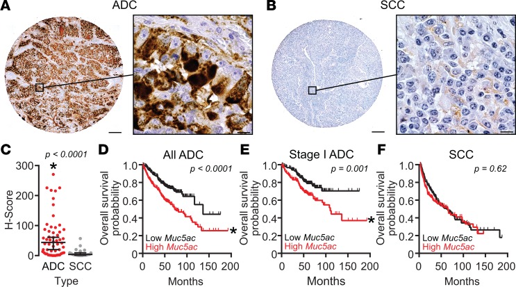
Figure 1. MUC5AC production is associated with human lung ADC and poor survival.
(A–C) MUC5AC protein levels were analyzed immunohistochemically using human tissue arrays. Representative images of MUC5AC immunostaining in adenocarcinoma (ADC) (A) and squamous cell carcinoma (SCC) (B) are shown. Scale bars: 200 μm (low magnification) and 10 μm (high magnification). (C) MUC5AC H-scores for ADC (n = 53) and adenosquamous (n = 3) (combined and included with ADC, red circles) were compared with SCC (n = 21, gray circles). MUC5AC levels were significantly higher in ADC tissues. Data are individual values with lines depicting medians ± 95% CIs. Significance was determined by the Wilcoxon rank sum test (61). P < 0.05. (D–F) Effects of MUC5AC expression on survival in ADC and SCC were tested using KM Plotter (http://kmplot.com/analysis/). MUC5AC expression was stratified to define high- (red) and low-expression (black) groups by median transcript levels. Kaplan-Meier survival plot and hazard ratio calculations were compared by multivariate log-rank analyses with sex and stage as covariates; P < 0.05. While sex was not significant, stage was strongly significant for ADC (log-rank P < 1 × 10–14). Significant effects of high MUC5AC gene expression were observed for all human lung ADC (D, n = 534, log-rank P < 0.0001) and for stage I ADC (E, n = 370, log-rank P = 0.001) samples. No differences were observed in SCC (F, n = 315, log-rank P = 0.62). For all groups in D–F, calculated hazard ratios were 2.0 (1.5–2.7) for all ADC, 1.9 (1.3–2.8) for stage I ADC, and 1.1 (0.8–1.5) for SCC.

