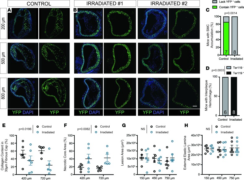
Figure 2. SMCs fail to invest within brachiocephalic atherosclerotic lesions following lethal irradiation and BMT; these changes are associated with decreased indices of lesion stability.
SMC-YFP, Apoe–/– mice received 1,200-cGy whole-body radiation and BMT followed by 18 weeks of WD to induce atherosclerosis lesion development. (A) High-resolution confocal imaging shows consistent YFP+ cell accumulation in brachiocephalic (BCA) lesions of control mice. (B) Lethally irradiated animals show loss of YFP+ cell accumulation in BCA lesions at 3 locations past the aortic arch. (C) Fisher’s exact test quantifying the percentage of control and irradiated animals that demonstrate YFP+ cell accumulation in BCA lesions. (D) Fisher’s exact test quantifying the percentage of animals with intraplaque hemorrhage in at least 1 of 3 locations along the BCA. (E) The percentage of collagen pixels in the 30-μm lesion cap area was significantly decreased after lethal irradiation. (F) The necrotic core area was significantly increased in BCA lesions after lethal irradiation. (G and H) Lesion area (G) and external elastic lamina area (H) were not changed between control and radiated animals E–H. Data were assessed by 2-way ANOVA. Data represent mean ± SEM. Sample number is indicated in the graph. Scale bar: 100 μm.

