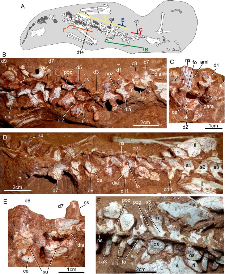
Figure 21. Vertebral column of Caipirasuchus mineirus, CPPLIP 1463.
Skeleton indicating the position of the illustrated vertebrae (A). Last cervical (c7–c8) to ninth dorsal (d9) vertebrae as positioned in the block (B). Detail of first (d1) and second (d2) dorsal vertebrae in right lateral view (C). Fourth (d4) to third sacral (s3) vertebrae as positioned in the block (D). Detail of partial sixth (d6) and seventh (d7) vertebrae in left lateral view (E). Detail of sacral and first caudal vertebrae in dorsal view (F). Abbreviations: aml, anterior medial lamina; c, cervical vertebra; ca, caudal vertebra; ce, vertebral centrum; d, dorsal vertebra; dia, diapophysis; fo, fossa; il, ilium; ns, neural spine; os, osteoderm; para, parapophysis; pml, posterior medial lamina; poz, postzygapophysis; prz, prezygapophysis; ri, rib; s, sacral vertebra; su, neural suture, vk, ventral keel.

