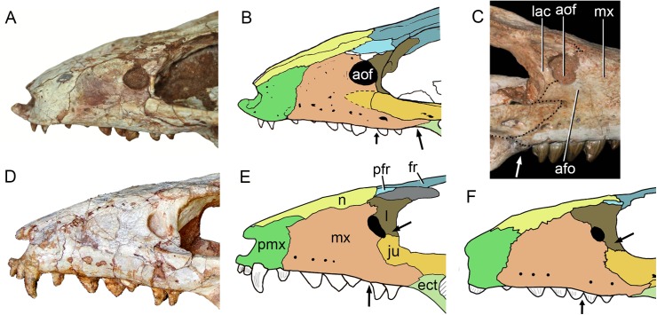
Figure 31. Comparison of the snout in Caipirasuchus spp. in lateral view.
Photograph of Caipirasuchus mineirus (holotype, CPPLIP 1463) (A) with schematic drawing (B). Photograph of Caipirasuchus stenognathus (holotype, MZSP-PV 139) (C). Photograph of Caipirasuchus paulistanus (holotype, MPMA 67-0001/00) (D) with schematic drawing (E). Schematic drawing of Caipirasuchus montealtensis (holotype, MPMA 15-0001/9) (F). Arrows indicate main differences in this portion of the skull. B was modified from Pol et al. (2014). Abbreviations: afo, antorbital fossa; aof, antorbital fenestra; ect, ectopterygoid; fr, frontal; ju, jugal; lac, lacrimal; mx, maxilla; n, nasal; pfr, prefrontal; pmx, premaxilla.

