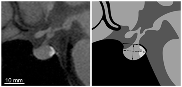
Fig. 1.

Left: Midsagittal T1-weighted MR image of the sella turcica and pituitary gland from a control subject. Right: Drawing showing the dimensions that were measured to determine the opening (solid line), depth (dotted line), and length (dashed line) of the sella turcica. In this example, the sellar area = 70 mm2, opening = 8.4 mm, length = 11.4 mm, depth = 7.6 mm, and pituitary area = 64 mm2.
