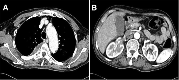
Fig. 3.
Contrast-enhanced computed tomography after 1 month of treatment. a Contrast-enhanced chest computed tomography shows reduction in size of the pleural mass. b Contrast-enhanced abdominopelvic computed tomography shows decreased infiltration around the right renal pelvis (longest diameter, 31 mm)

