Figure 1.
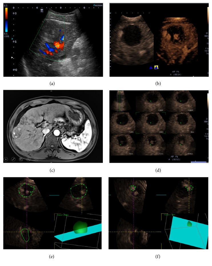
Residual tumor after radiofrequency ablation (RFA). Conventional ultrasound image in a 65-year-old man showed a 45 mm sized homogeneous hypoechoic lesion in the right hepatic lobe. No color signal can be detected inside the lesion by color Doppler flow imaging (CDFI) (a). There was a small enhanced area along the border of the lesion on dynamic 3D-CEUS (right) other than 2D-CEUS (left) images (b). Contrast MR imaging showed confirmed RT inside the lesion with local enhancement (c). The “multislice” display mode showed the RT area on several slices of 3D-CEUS image (d). The contour of the whole lesion was depicted manually and its corresponding volume was calculated automatically (e). The volume of the 3 nodular RT area can also be calculated. The residual proportion added up to 13.4% (f).

