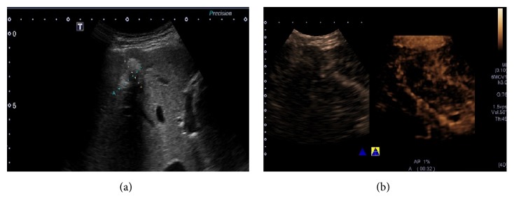
Figure 3.
Presence of attenuation due to RFA treatment. B mode ultrasound image in a 71-year-old man showed a 25 mm sized lesion treated by RFA in the right lobe of the liver. It had posterior attenuation caused by scars or necrosis (a). Its posterior border cannot be demonstrated by 2D-CEUS (left), whereas it can be shown on dynamic 3D-CEUS. The relationship of the lesion and the posterior adjacent large vessels can also be shown (b).

