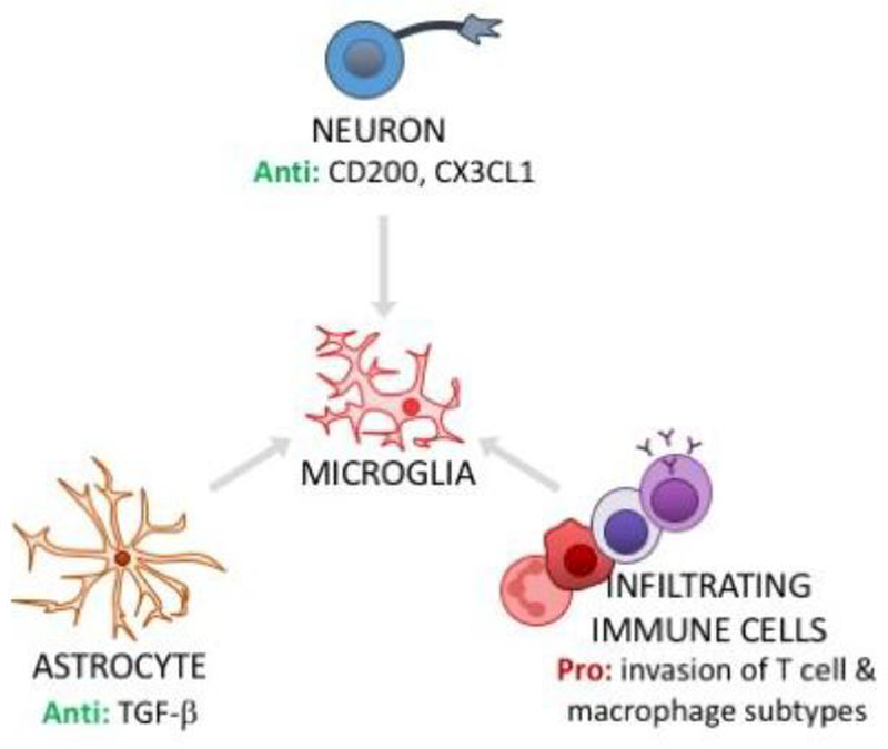
Figure 3. Microglial phenotype is regulated by other cells in the CNS.
In healthy CNS, steady-state microglial quiescence is maintained by signaling with neurons (neuron CD200L to microglia CD200R; neuron CX3CL1 to microglia CX3CR1) and astrocytes (e.g., TGF-β signaling limits microglial activation). Dysregulation of these cell-cell interactions can contribute to microglial priming. In addition, hematogenous immune cells - which typically have no/very limited access to the CNS - can invade CNS tissue in stress and aging. Pro-inflammatory subtypes of infiltrating cells, such as macrophages, can signal to microglia to elicit priming and/or activation. Anti = anti-inflammatory actions; Pro = pro-inflammatory actions.

