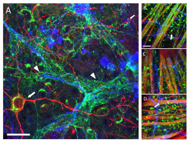
Figure 1.
Changes in GM1 ganglioside in the DBA2/J and DBA/2-Gpnmb+ retina as determined by cholera toxin-B (CTB) binding. Panels show CTB (green, AlexaFluor-488) labeling in the retina, with β-Tubulin III (red, AlexaFluor-594) and GFAP (blue, AlexaFluor-647) to label retinal ganglion cells (RGCs) and astrocytes, respectively. A. A 13 month-old DBA/2J retina with CTB-positive astrocytes (arrowheads). Arrow points to a β-Tubulin III-positive retinal ganglion cell. B. A 3 month-old DBA/2J retina showing RGC axon fascicles (bundles of green) and no CTB-positive astrocytes. Arrows point to CTB-positive RGCs. C. A 3 month-old DBA/2-Gpnmb+ retina, also with CTB-positive RGC axon fascicles and GFAP-positive astrocytes. D. A 13 month-old DBA/2-Gpnmb+ retina that appears similar to the 3-month-old retina. Arrows point to CTB-positive RGCs. The GFAP-positive astrocytes do not have CTB labeling. The astrocytic CTB labeling occurs only in glaucomatous (panel A), degenerating retina. Scale bar = 20 Jm

