
Figure 2.
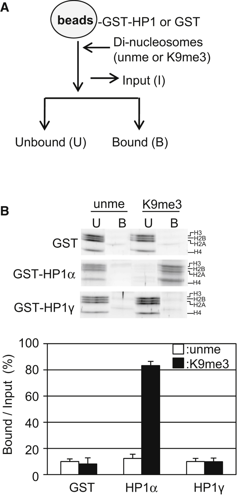
Biochemical assay of HP1 binding to dinucleosomes with 25-bp linker DNA. (A) A schematic illustration of the pull-down assay is given. The number of dinucleosomes bound to GSH Sepharose on which GST-HP1 or GST were anchored were analyzed. (B) HP1 isoform-specific dinucleosome binding activity is shown. Dinucleosomes reconstituted with unmethylated H3 (unme) or H3K9me3(K9me3) were mixed with the beads, and the unbound (U) and bound (B) fractions were separated and then subjected to SDS-PAGE (upper panel). SDS-PAGE data are taken from the whole gel image shown in Fig. S11. The amounts of each histone in the bound fractions over input (%) are shown as mean ± standard error (n = 3) (lower panel).