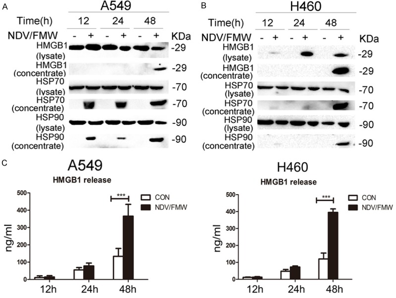
Figure 3.

Release of immunologic signals upon NDV infection. A, B. Cell-free supernatants (concentrated) and whole cell lysates were collected from A549 and H460 cells infected with NDV/FMW (MOI = 10) for distinct periods of 12, 24 and 48 h. Immunoblot (IB) analysis of HMGB1 and HSP70/90 in either whole cell lysates or concentrated supernatants of A549 and H460 cells. Data shown are representative of two independent experiments. C. Enzyme-linked immunosorbent (ELISA) detection of HMGB1 release in NDV/FMW cell supernatants compared to uninfected control cells (***P<0.001, n = 4).
