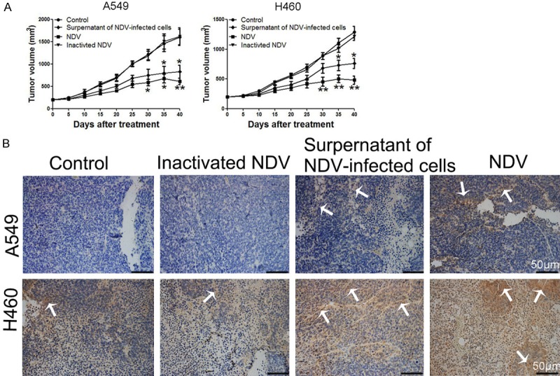
Figure 6.

Tumor growth in response to supernatants from NDV-infected lung cancer cells. A. A549 and H460 cells were injected subcutaneously into the right flanks of mice to establish tumors. When tumors reached approximately 200 mm3, mice received an intratumoral injection of either PBS, the concentrated cell-free supernatants of NDV/FMW (50 μl), NDV or inactivated NDV every three days. Tumor volumes were measured at 5-day intervals for 40 days after injections and expressed as the Mean ± SD (n = 10) and represented as tumor volume-time curves to show any differences in tumor regression (*P<0.05, **P<0.01). B. Immunohistochemistry assay for expressions of CRT protein in four groups of mice model, arrowheads indicate positive area. Scale bar = 50 μm.
