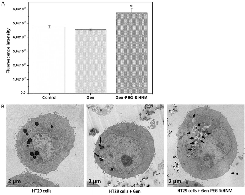
Figure 9.

A. Autophagy’s activation in HT29 cells after 24 h of incubation with genistein (Gen) and Genistein-PEGylated silica hybrid nanomaterials (Gen-PEG-HNM) at IC50 (24.3 μM). *indicate statistical difference (P < 0.05) between treatments and control group analyzed by Dunnet’s test. B. Autophagic structural analysis by TEM in HT29 cells exposed to 23.43 µM of free or encapsulated Gen for 24 h. Arrows show the presence of autolysosomes formed after cells were exposed to treatments for 24 h. Untreated HT29 cells were used as control.
