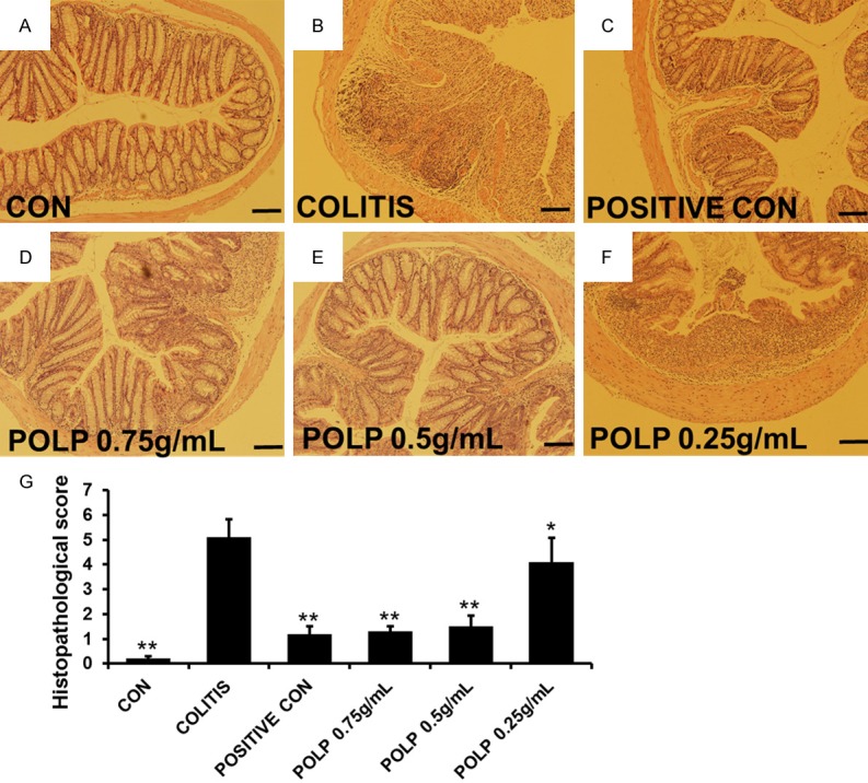
Figure 3.

Histologic characteristics of each group, magnification ×100. A. Normal colonic mucosa with regularly formed colonic folds covered by intact mucosa. B. The histologic characteristics of colitis group. Inflammatory cell completely infiltrated to mucosa and submucosa. C. The histologic characteristics of sulfasalazine control group. D. The histologic characteristics of POLP group with a concentration of 0.75 g/mL. E. The histologic characteristics of POLP group with a concentration of 0.5 g/mL. F. The histologic characteristics of POLP group with a concentration of 0.25 g/mL. G. The sum of histological score obtained from blinded histopathological analysis. Error bars represented means ± SEM of n=3, *P<0.05 and **P<0.01 compared with colitis.
