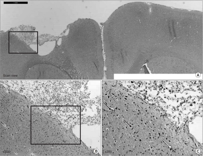
Fig. 8.
(A) The hematoxylin-eosin (H&E) staining showed the traumatic injured area of the affected hemisphere. (B) Note lots of dead cells that have a pyknotic nucleus throughout the brain parenchyme and large cavity that involved the cortex at the site of injury (H&E, ×100); and (C) the pyknotic nucleus-dead cells (black arrow) and vacuolated nucleus (blue arrow) in the brain cell after the traumatic brain injury (H&E, ×200).

