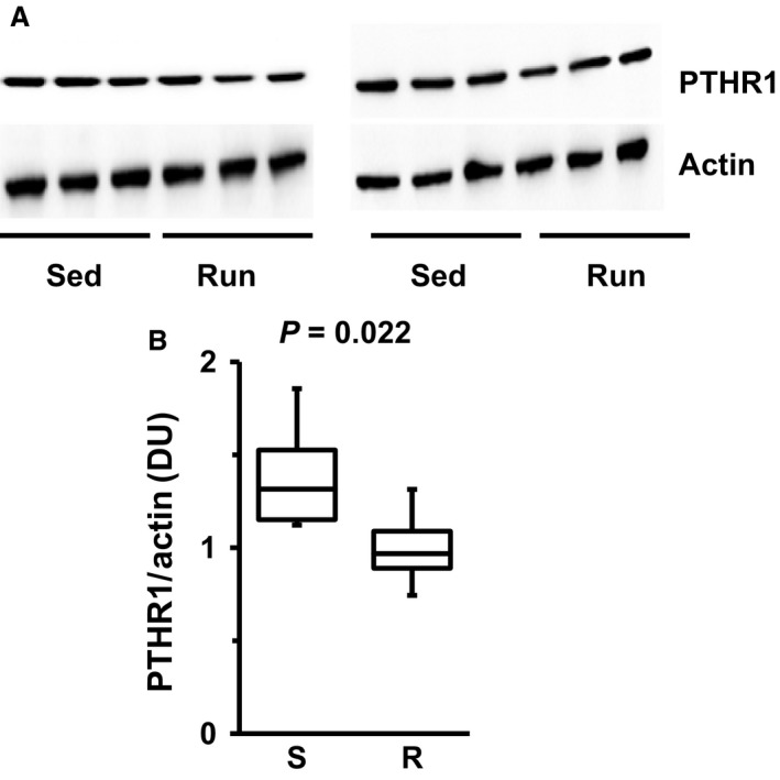
Figure 4.

Western blots indicating renal protein expression of PTHR1 in six SHRs after 10 months of exercise. (A) Original Western Blots for PTHR1 and actin, taken as a loading control. (B) Densitometry of the blots and quantification as densitometric units (DU) of PTHR1 normalized to actin. Data are means for time profiles and box plots representing the 25th, 75th quartile, and median, with whiskers representing the lowest and highest values (range). Data are from n = 6 rats. Exact p‐values are given.
