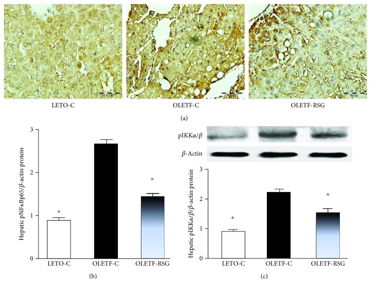
Figure 5.
Representative images showing immunohistochemical staining (a, ×200) and expression level (b) of hepatic phosphorylated NF-κBp65 (pNF-κBp65) protein and hepatic pIKK-α/β protein expression by Western blot (c) in LETO-C, OLETF-C, and OLETF-RSG groups. The result in the LETO-C group was arbitrarily assigned a value of 1. Data are means ± SD (n = 8 each group) versus OLETF group (∗P < 0.05).

