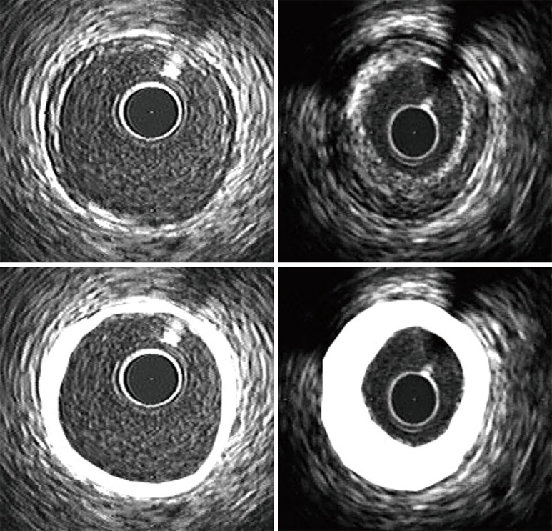
Figure 1.
Illustrative example of differences in remodelling between a female subject with symptomatic ischemia in the absence (left panels) or presence (right panels) of obstructive disease on coronary angiography. Location of atherosclerotic plaque in cross-sectional images is depicted by shading in the lower panels. A lack of compensatory expansion of the outer vessel border in the presence of a greater plaque burden results in a smaller lumen, which would be detected as an abnormality on angiography.

