
Figure 4.
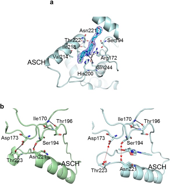
Second gefitinib binding site. a) Location of the second gefitinib binding site with an overlay of the 2 F o−F c electron density map (1σ) of gefitinib. b) Structures of GAK_1 (green ribbon structure; left panel) and GAK_2 (cyan ribbon structure; right panel) around the second binding site. A gefitinib molecule bound to GAK_2 is shown as a stick model.