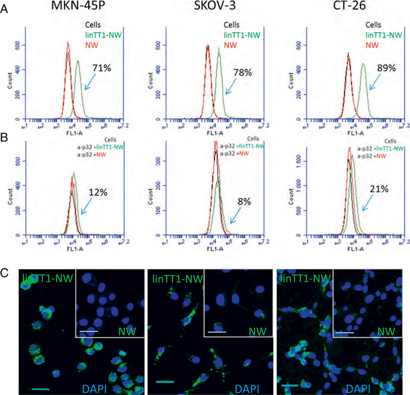
Fig. 2.

LinTT1-NWs bind to peritoneal carcinomatosis cell lines in a p32-dependent manner. (A) Flow cytometry of MKN-45P, SKOV-3 and CT-26 cells incubated with linTT1-NWs and control FAM labeled NWs. Cells in suspension were incubated with NWs (at 30 μg/ml Fe) for 1 h followed by washes, and flow cytometry analysis. Green line: cells incubated with linTT1- NWs; red line: cells incubated with NWs; black line: cells without NW incubation. (B) Anti-p32 antibody inhibition of NW binding to MKN-45P, SKOV-3 and CT-26 cells. Suspended cells were pre-incubated with 20 μg/ml of p32 antibody, followed by NW incubation for 1 h, washes and flow cytometry. The labeling colour scheme is the same as in A. (C) Fluorescence confocal imaging of cultured adherent MKN-45P, CT-26 and SKOV-3 cells incubated with linTT1-NWs or non-targeted NWs for 3h. Green: NWs; blue: DAPI. Scale bars: 30 μm. (For interpretation of the references to colour in this figure legend, the reader is referred to the web version of this article.)
