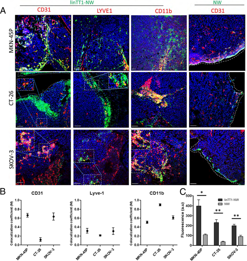
Fig. 5.

Targeted NW homing in different models of peritoneal carcinomatosis. (A) Confocal Imaging of tumor sections. Mice bearing MKN-45P (upper panel), CT-26 (middle panel) and SKOV-3 (lower panel) tumors were Injected with linTT1-NWs (5 mg iron/kg). Tissues were collected 5 h after IP injection of NWs, and cryosections of tumor tissue were stained with antibodies against CD31 (blood vessels). LYVE-1 (lymphatic vessels) and CD11b (macrophages). Green: NWs; Red: CD31, LYVE-1 or CD11b. Blue: DAPI. Scale bars: 100 μm. (B) Colocalization analysis of linTT1-NWs with CD31, LYVE-1 and CDllb in MKN-45P, CT-26 and SKOV-3 tumor models based on Manders (M) coefficient [39] 0 = no colocalization; 1 = perfect colocalization. Analysis was performed by ImageJ software. Error bars, mean ± SEM (C) Quantification of green fluorescence intensity in the confocal images of tissue sections prepared from MKN-45P, CT-26 and SKOV-3 tumors. Representative fields from multiple sections representing tumors from 3 mice per group are shown. Analysis by Image J, N > 3 mice; Statistical analysis: Student’s t-test; error bars: mean ± SEM; **p < 0.01, *p < 0.05. (For interpretation of the references to colour in this figure legend, the reader is referred to the web version of this article.)
