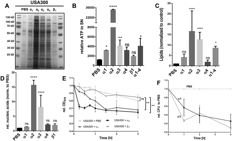
Figure 4: Exogenous supplied PSMα peptides enhance cell leakage and ECP.
(A) SDS-PAGE of the extracellular proteins of USA300, treated with PSMα1, α2, α3, α4 and β1. (B) Extracellular ATP levels of PBS washed USA300 cells incubated with PBS, α1, α2 α3, α4 and β1 after incubation for 4h. (C) Relative amount of membrane lipids in the supernatant of PSM treated USA300 cells after 4 h incubation of washed cells in the mid exponential growth phase. (D) Relative extracellular nucleic acids, normalized to PBS, after treatment of USA300 with synthetic PSM peptides. (E) Relative OD of mid exponential growth phase cells of USA300, WT* and WT*Δpmt resuspended in PBS and monitored for 3 h in PBS, PBS + α1, PBS + α3 and PBS + β1. (F) Relative CFU of PBS compared to PSMα1 and PSMα3 treated USA300 cells for a period of 4 h. Representative data from at least two independent experiments are shown. For all graphs, each data point is the mean value ± SD (n = 3 for BCD and n = 2 for EF) *p < 0.05; **p < 0.01; ***p < 0.001; and ****p < 0.0001, by one-way ANOVA with Bonferroni posttest for BCD and unpaired t-test for EF.

