Figure 1.
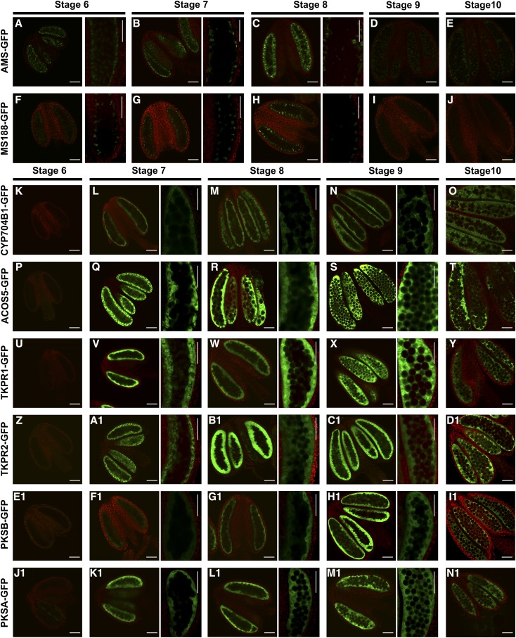
Protein localization of AMS-GFP, MS188-GFP, and sporopollenin synthesis protein-GFPs. Confocal images of the fluorescence of the AMS-GFP, MS188-GFP, CYP704B1-GFP, ACOS5-GFP, TKPR1-GFP, TKPR2-GFP, PKSB-GFP, and PKSA-GFP fusion proteins from stages 6 through 10. GFP expression (530 nm) is shown in the green channel, while chlorophyll autofluorescence (>560 nm) is shown in the red channel. A to C and F to H, The fluorescence of AMS-GFP and MS188-GFP at stages 6 through 8. Right, high-magnification images of GFP localization. D, E, I, and J, The fluorescence of AMS-GFP and MS188-GFP at stages 9 and 10. K, P, U, Z, E1, and J1, The fluorescence of CYP704B1-GFP, ACOS5-GFP, TKPR1-GFP, TKPR2-GFP, PKSB-GFP, and PKSA-GFP at stage 6. L to N, Q to S, V to X, A1 to C1, F1 to H1, and K1 to M1, The fluorescence of CYP704B1-GFP, ACOS5-GFP, TKPR1-GFP, TKPR2-GFP, PKSB-GFP, and PKSA-GFP at stages 7 to 9. Right, high-magnification images. O, T, Y, D1, I1, and N1, The fluorescence of CYP704B1-GFP, ACOS5-GFP, TKPR1-GFP, TKPR2-GFP, PKSB-GFP, and PKSA-GFP at stage 10. Scale bars, 50 μm.

