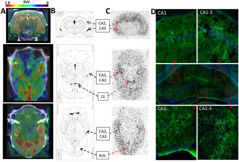
Figure 3.
(A) PET−CT−MR coronal and axial images 20 min after iv administration of 2-[18F]BzAHA, demonstrating increased 2-[18F]BzAHA-derived accumulation within regions of the brain corresponding to the hippocampus (CA1−CA3), lateral septal nucleus (LS), and nucleus accumbens (Acb). (B) Maps of the brain corresponding to the PET images. (C) Autoradiography performed for matching sections of the brain at 20 min after iv administration of 2-[18F]BzAHA demonstrating increased radioactivity within regions of the hippocampus, nucleus accumbens, and lateral septal nucleus. (D) Immunofluorescence of corresponding slices of the brain demonstrated increased levels of SIRT1 expression in the hippocampus (Alexafluor488-conjugated anti-SIRT1, green), especially CA2 and CA3, overlaid on DAPI nuclear stain (blue).

