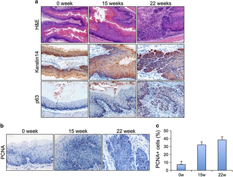
Figure 1.
Mice treated with carcinogen developed dysplasia and ESCC. (a) H&E staining and K14 and p63 IHC staining of normal esophageal epithelium, dysplasia and SCC. (b) PCNA IHC for cell proliferation in normal esophageal epithelium, dysplasia and SCC. (c) Quantification of cell proliferation in normal esophageal epithelium, dysplasia and SCC. H&E, hematoxylin and eosin. IHC, immunohistochemical.

