Figure 1.

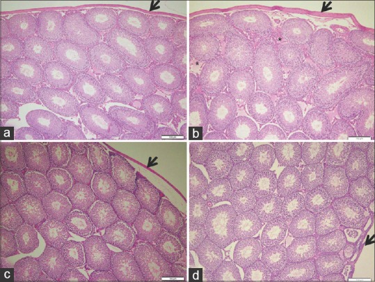
Photomicrographs of rat testis showing (a) control, normal structure of seminiferous tubules separated by interstitial tissue. (b) ochratoxin A, proliferation of interstitial tissue (*) and apparent thickened tunica albuginea capsule (thick arrow). (c) Ajwa dates, typical tubular structure with tightly packed seminiferous tubules and well-defined basal lamina. (d) Ajwa dates and ochratoxin A, normal testis structure with compacted seminiferous tubules. Note decreased capsule thickness in Ajwa dates groups (thick arrow) (H and E ×100)
