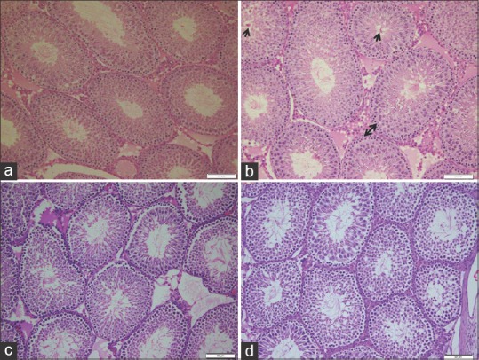
Figure 2.

Photomicrographs of rat seminiferous tubules showing (a) control (b) ochratoxin A, disorganized spermatogenic cells with a marked increase of spermatogonia (two heads arrow). Note increased eosinophilic material and clumps of coagulated sperms inside the lumen (short arrow). (c) Ajwa dates, complete spermatogenic series and a well-defined basal lamina. Note the apparent increased number of sperm count inside the lumen. (d) Ajwa date and ochratoxin A, seminiferous tubules with complete stages of spermatogenic cells and apparent increased number of sperms (H and E, ×200)
