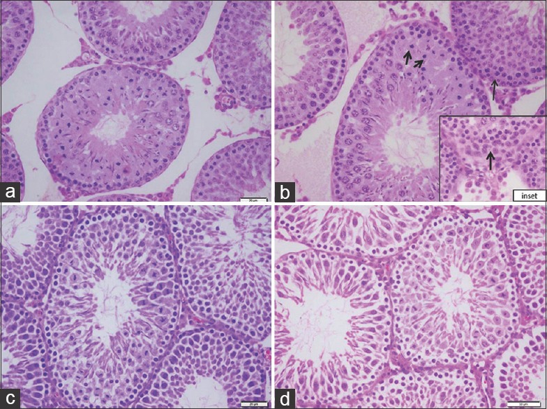
Figure 3.

Higher magnification of seminiferous tubules showing (a) control (b) ochratoxin A, degenerative changes with loss of the spermatogenic series (arrows). (inset; base of the same tubule shows degeneration of the basement membrane and proliferation of Sertoli cells (arrow). (c) Ajwa dates, seminiferous tubules with all spermatogenic series. (d) Ajwa dates and ochratoxin A, remarkable improvement, closely packed tubular structure with primary spermatocytes, early and late spermatids as well as a large number of sperms inside the lumen (H and E, ×400)
