Figure 1.

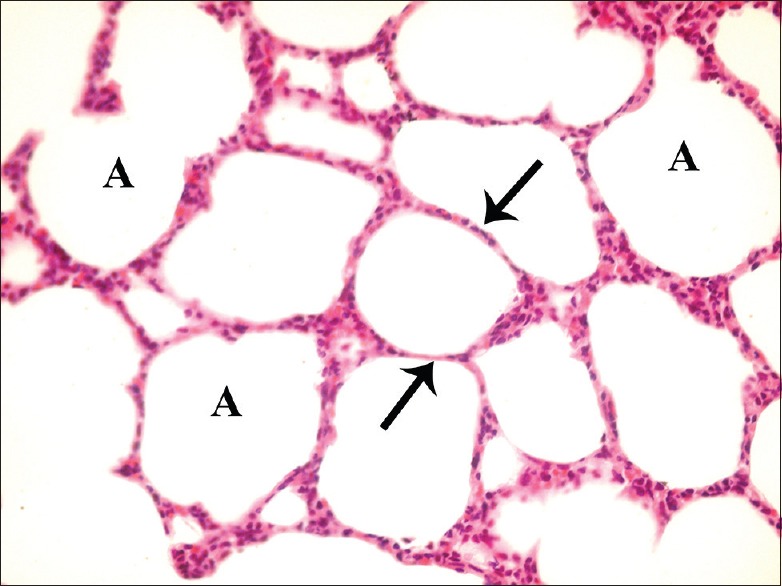
A photomicrograph of a lung section of control group showing normal lung architecture with spongy structure, normal clear alveoli (A), and thin inter-alveolar septa (arrow) (H and E, ×400)

A photomicrograph of a lung section of control group showing normal lung architecture with spongy structure, normal clear alveoli (A), and thin inter-alveolar septa (arrow) (H and E, ×400)