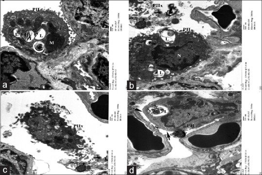
Figure 9.

Electron micrographs of ultrathin section of experimental group I showing (a) type II pneumocyte (PII) lining the alveolar cavity with multiple empty lamellar bodies (L) and megamitochondria(M). (b) Nucleus of pneumocyte type II appears indented with electron dense chromatin. (c)Some pneumocyte type II exfoliated in the lumen (PIIx) and other showing blunt surface without microvilli (arrow). Notice congested blood capillaries (bc) (d) and collagen fibers (Co) (a)
