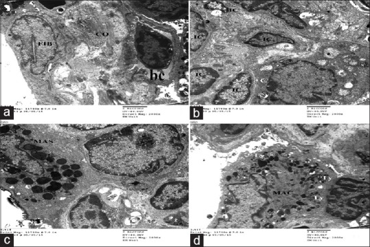
Figure 11.

Electron micrographs of ultrathin section of experimental group II showing thick inter alveolar septa with deposition of excess collagen fibers associated with active fibroblast (FIB) with euochromatic nucleus (a) and apparent increased numbers of interstitial cells (IC) (b), different inflammatory cells like mast cells (MAS), and active macrophages (MAC) with excess electron dense bodies and secondary lysosome(Ly) (c&d). Notice blood vessels (bc) (a)
